More precise cancer diagnosis thanks to 3D computed tomography
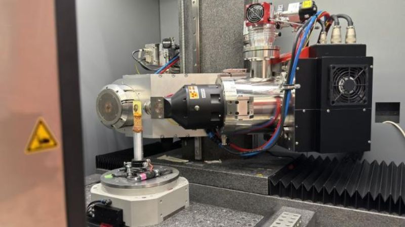
The new 3D tissue analysis of thyroid tumors is still unfamiliar to pathologists. Instead of cutting the removed tissue into thin slices and viewing them two-dimensionally under the microscope as before, they can now examine the entire tissue sample virtually on the screen and rotate it as required to identify pathological changes. This has been made possible by so-called non-invasive histopathological 3D imaging. “The special thing about this method is that it can analyze complete biopsy blocks of a tumor in three dimensions in a short time without changing or destroying the tissue. This means that the sample can still be used for further molecular biological examinations,” says Robert Zboray, group leader at Empa's Center for X-ray Analysis, who developed this technology.
Personalized treatment
Together with pathologists from the University of Bern, Zboray was able to demonstrate that his new method can detect clinically relevant tissue characteristics in thyroid tumors. X-ray phase-contrast micro-computed tomography (micro-CT) makes even the smallest differences in soft tissue visible. These three-dimensional images of tissue samples are then analyzed using machine learning. The Empa researcher hopes that this will enable pathologists to make more precise diagnoses and prognoses. The greatest challenge is to treat patients as individually as possible - in other words, to avoid overtreatment of low-risk tumors and at the same time to treat and monitor patients with a higher risk appropriately.
Around 300 million people worldwide are affected by thyroid cancer. However, tumor characteristics often differ from patient to patient. These measurable biochemical and molecular characteristics of a tumor are known as biomarkers. They help to detect cancer at an early stage or indicate how aggressively a tumor can grow and which therapy it may respond to.
